Brachycephalic, or flat-faced, pets remain popular in America despite their high prevalence of morbidities. During 2019, the 4th and 5th most popular dog breeds nationwide were the French Bulldog and (English) Bulldog, respectively, while the most popular pedigreed cat breed was the Persian. The French Bulldog has become the most popular breed in many major cities, where they are currently ubiquitous. With no signs of this popularity waning, veterinary professionals must be familiar with the idiosyncrasies particular to the brachycephalic pet and prepared to deal with the associated complications.
Anesthesia of brachycephalic pets carries an increased risk of airway-related complications as structural abnormalities increase airway resistance causing some degree of upper airway obstruction. A primary consideration in brachycephalic anesthesia is preventing, and being prepared to manage, upper airway obstruction, especially at the times of highest risk, which are: 1) following premedication, 2) at anesthetic induction, 3) upon extubation, and 4) during any period of increased stress.
Common Pathology
Brachycephalic breeds are predisposed to abnormalities in a wide variety of body systems, with respiratory, cardiovascular, and gastrointestinal pathology most relevant to anesthesia. The high incidence of obesity in brachycephalic breeds can also compromise respiratory and cardiovascular function during anesthesia.
Respiratory
Excessive pharyngeal soft tissue and reduced airway size are common in brachycephalic animals, resulting in increased airway resistance and effort required to breathe. Both primary abnormalities and secondary changes in airway anatomy (see Table 1, Figures 1 and 2) impair respiratory efficiency.
Table 1: Common primary conformational abnormalities and secondary changes in the airway anatomy of brachycephalic breeds.
Cardiovascular
High vagal tone is commonly present in brachycephalic animals, increasing the risk of bradycardia during anesthesia, especially with the use of multiple cardiovascular depressant medications.
Gastrointestinal
Vomiting, regurgitation, gastroesophageal reflux and other commonly reported gastrointestinal abnormalities increase the risk of aspiration pneumonia and contribute to the importance of rapidly securing airway control following induction of anesthesia of brachycephalic patients. Gastrointestinal pathology has been detected in brachycephalic patients that have never exhibited related clinical signs, suggesting that even the “healthy” members of this population may be at increased risk of gastrointestinal complications during anesthesia.
Patient Assessment
History
One study demonstrated that a majority of surveyed Pug, French Bulldog, and Bulldog owners assessed their dogs as very healthy, at odds with the high prevalence of various abnormalities in their pets. Owners may not be aware that respiratory stridor or stertor, exercise intolerance, and heat intolerance are abnormal, and could be associated with increased risk during anesthesia.
The presence and severity of respiratory compromise during every day activities may indicate the extent of a patient’s airway abnormalities. A history of ptyalism, vomiting or regurgitation suggest increased likelihood of either pre-existing aspiration pneumonia or development of this post-operatively.
Physical examination
Unless a physical examination is likely to produce a respiratory crisis, a full examination of the patient, including an assessment of nares conformation, respiratory pattern, mucous membrane color, body condition scoring, and temperature, should be performed. Further diagnostics or stabilization may be indicated prior to sedation or anesthesia, depending on abnormalities in respiratory status, conformation and/or function.
Further diagnostics
Hematology, serum biochemistry and urinalysis may be considered to assist in diagnosing and ruling out comorbidities. Thoracic radiographs may be indicated, to assess for hypoplastic trachea, pre-existing aspiration pneumonia, and other abnormalities, especially in animals with a recent history of vomiting or regurgitation. Radiographs also provide a baseline, which may assist in assessing any post-operative aspiration pneumonia.
Anesthetic Considerations
Anesthetic plans should be individualized for each patient and consider their signalment and status, the intended procedure, clinician familiarity with medications, and up-to-date anesthesia guidelines. See Table 2 for a Quick Reference Guide to Anesthesia in Brachycephalic Dogs and Cats.
At-Home
Before the patient arrives for a procedure, pet owners should be provided clear instructions for preparation, including withdrawal of food and water and any medications to be administered. The 2020 AAHA Anesthesia and Monitoring Guidelines for Dogs and Cats recommend withholding food for 4 – 6 hours, and not withholding water at all in healthy animals over eight weeks of age. For patients with a history or increased risk of regurgitation it is recommended that both food and water be withheld for 6 – 12 hours, although a small amount of wet food may be considered 4 – 6 hours prior to induction.
Antiemetic medications can reduce the risk of vomiting and aspiration pneumonia when used appropriately. Oral maropitant, administered two hours prior to premedication, has been demonstrated to reduce the incidence of vomiting associated with hydromorphone in dogs and with morphine and dexmedetomidine in cats. Oral maropitant 18 hours prior to premedication also reduced vomiting and retching in cats suggesting that administration the evening prior to admission is also appropriate and may be easier for some pet owners. If at-home oral administration of maropitant is not possible, subcutaneous injection should occur at least 60 minutes prior to premedication with emetic opioids and/or sedatives.
At-home administration of oral anxiolytics can significantly reduce the stress associated with transport to the veterinary clinic. Trazodone and/or gabapentin are often administered to dogs for this purpose while gabapentin alone is commonly administered to cats. These medications should be administered two to three hours prior to admission.
In-Clinic Preparation
Anesthesia of a brachycephalic pet requires careful monitoring and support from before premedication until total recovery from anesthesia. Preparation to induce and intubate ahead of schedule is essential as upper airway obstruction may occur with little warning.
All efforts should be made to minimize patient stress once at the veterinary clinic, ideally using minimal restraint, keeping the animal in a cool area with some ability to move around, and avoiding delays prior to the procedure. Placement of topical local anesthetic over intended venipuncture and catheter insertion sites on presentation may lead to less stressful sample collection and intravenous catheter placement.
Premedication
Sedation improves ventilation, due to slower inspiratory flow and reduced agitation, but increases the risk of airway obstruction due to pharyngeal muscle relaxation and blunted compensatory responses. Short-acting or reversible medications are preferable to longer-acting, non-reversible medications and if a patient is quiet enough to receive only an opioid premedicant, one without emetic properties is ideal.
Following premedication, patients should be closely monitored for increased upper respiratory sounds or other signs of impending obstruction. Intravenous catheterization should occur as soon as possible, without causing undue stress, to facilitate intervention with drugs and/or fluids if airway function deteriorates.
The use of sedatives, usually combined with an opioid, may be required in all but the quietest patients. The benefits of reduced stress and easier handling are balanced against the risks of drug-induced side effects for each individual patient. The successful administration of low doses of acepromazine are frequently reported despite its long duration of action. Alpha 2-agonists are somewhat controversial as the benefits of reliable, potent sedation bring associated risks of marked upper airway relaxation, vomiting and exacerbation of bradycardia; therefore, they should be used with caution and at low doses.
Butorphanol’s antitussive properties may increase tolerance of intubation post-operatively so it is a good opioid option for non-painful procedures. For painful procedures, methadone or buprenorphine are unlikely to induce vomiting so may be preferable to hydromorphone or morphine, especially without pre-administration of an antiemetic. An anticholinergic may be used to antagonize high vagal tone and decrease the risk of bradycardia, especially if pure μ-agonist opioids are administered.
Multimodal analgesia should be considered in every patient, including locoregional anesthesia during the procedure whenever possible. Non-steroidal anti-inflammatory drugs should be reserved until the patient has fully recovered from anesthesia in case corticosteroids are required to treat airway abnormalities post-operatively.
Further gastrointestinal support may be provided with a proton pump inhibitor, antiemetic, and/or prokinetic, according to the patient’s condition, procedure to be performed, and other medications administered.
Pre-oxygenation
Provision of supplemental, medical grade oxygen prior to induction of anesthesia increases the time before oxygen desaturation occurs in a patient experiencing apnea or respiratory obstruction. Delivery of 100% oxygen via a tight-fitting mask (Figure 3) for three minutes is preferable, but the mask may increase obstruction risk in stressed animals. In patients that will not tolerate a mask, provision of flow-by oxygen is likely to increase PaO2 compared to breathing room air.
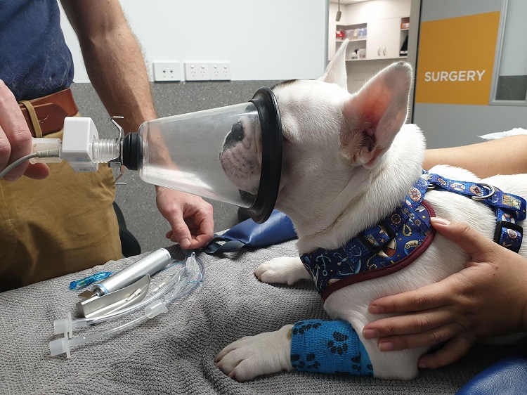
Figure 3: Pre-oxygenation with 100% oxygen via face mask prolongs the onset of desaturation if apnea or respiratory obstruction occur
Anesthetic Induction
Animals should be in sternal recumbency and flexion of the neck avoided to prevent airway collapse on induction. Recommended characteristics of an induction agent for brachycephalic animals include:
- Rapid onset of anesthesia, allowing an airway to be secured without delay
- Minimal disruption of cardiorespiratory parameters
- Short duration of action
- Rapid recovery of normal behavior (and airway control)
Alfaxan Multidose (alfaxalone) is an appropriate choice for anesthetic induction of brachycephalic patients, meeting each of the above criteria. Safe and effective alfaxalone use in brachycephalic dogs for laryngeal assessment and undergoing cesarean sections has been reported, including use for both induction and maintenance of anesthesia. See the Alfaxan Multidose Prescribing Summary for further information on dosing and administration of alfaxalone.
Other short-acting injectable anesthetics may also be appropriate, as may a co-induction technique. Co-inductions generally involve an initial small amount of primary induction agent (usually alfaxalone or propofol), followed by a secondary co-induction agent (commonly a benzodiazepine, sometimes ketamine) with the induction completed by more of the primary agent, to effect.
Endotracheal Intubation
Rapid, successful intubation may be assisted by:
- Ensuring adequate depth of anesthesia
- Extending the head with the neck fully straightened to avoid respiratory obstruction
- Passing bandage strips behind both pairs of canine teeth to hold the mouth open, assisting visualization without interference of excess labial skin
- Gently using a suction device to clear obstructing saliva and mucous from the upper airways without damaging vocal folds
- Using a laryngoscope with a long, curved blade and bright light source
- Preparing endotracheal tubes of different diameters as airways are often much smaller and less accessible than expected
- Using a tongue depressor to hold an elongated soft palate up
Once visualized, the larynx may be assessed for an elongated soft palate, everted laryngeal saccules and other pathology (Figure 4). Supplemental oxygen should be supplied immediately following intubation and respiration carefully monitored, with judicious ventilation performed, if required.
For further information on intubation, see the Think Anesthesia Endotracheal Intubation video.

Figure 4: Small airways with large amounts of soft tissue can make intubation of brachycephalic patients challenging
Anesthesia Maintenance and Monitoring
Inhalant anesthetics or partial/total intravenous anesthesia (PIVA or TIVA) with Alfaxan Multidose are appropriate for maintenance of anesthesia.
Respiratory monitoring including capnography and pulse oximetry allow insight into ventilatory efficiency throughout anesthesia and can guide the efficient use of manual or mechanical ventilation if required. Arterial blood gas analysis is recommended in more critical patients.
Cardiovascular monitoring should include arterial blood pressure monitoring (non-invasive in stable patients) and continuous electrocardiography. If excessive bradycardia occurs, and anesthetic depth is otherwise considered appropriate, an anticholinergic may be administered.
Eyes should be lubricated frequently, due to brachycephalic animals’ proclivity for exophthalmos, increasing the risk of corneal ulceration.
Recovery from Anesthesia
During recovery from anesthesia the patient should be placed in sternal recumbency with the head extended and tongue gently pulled rostrally. The observer assigned to monitor the higher risk brachycephalic patient should include in their monitoring pulse oximetry, respiratory character and body temperature, as hyperthermia may indicate airway obstruction. The endotracheal tube should remain in place for as long as the patient will tolerate it and oxygen saturation monitored both before and after extubation. Supplemental oxygen and equipment to re-anesthetize and intubate should be close at hand, in case of an emergency.
Pain scoring can be performed once the patient regains consciousness and any adjustments to the pain management plan made at this time.
Close monitoring of brachycephalic patients should continue well after extubation with owners encouraged to keep the animal quiet, in a cool place, following discharge.
When to Intervene During Recovery
Any evidence of post-operative airway obstruction, increasing body temperature and/or an SpO2 of less than 94% indicate the need for supplemental oxygen. Mild sedation may assist in patients that are beginning to show signs of respiratory distress.
If there is no improvement in the respiratory status of the patient within 10-12 minutes, or respiratory signs worsen, anesthesia and re-intubation should be performed. If laryngeal swelling is evident, continuing light anesthesia, possibly with mechanical ventilation for several hours may be necessary before attempting re-extubation.
Anti-inflammatory doses of a short-acting corticosteroid may be used to reduce edema of the airway soft tissue, if required. In cases of airway obstruction where an airway cannot be secured, an emergency tracheostomy and transtracheal oxygen are indicated.
Summary
Anesthesia of brachycephalic dogs and cats carries increased risk of airway obstruction and complications related to a variety of organ systems. Successful management of the brachycephalic anesthetic patient is achieved through careful planning, with a focus on minimizing stress, pre-oxygenation, vigilant monitoring, and preparedness to manage upper airway obstruction, bradycardia, and regurgitation. The highest risk periods are following premedication, following induction until intubation has occurred, and recovery, especially extubation.
Anesthetic plans should be made based on the patient’s status, procedure to be performed and clinician’s knowledge and experience with medications they have chosen. The use of short-acting injectable anesthetics that have a rapid onset of action, minimal cardiopulmonary depression, do not induce emesis and allow rapid recovery of laryngeal function upon cessation of anesthesia are recommended. Alfaxan Multidose meets these criteria and is suitable for induction of anesthesia, followed by maintenance with either an inhalant anesthetic or further alfaxalone.
Click here for a complete list of references cited in this article. This content is sponsored by Alfaxan and Think Anesthesia.